Macula Off, Next List
A macula-off detachment usually waits for the next routine list. What the first week of that wait costs, in eyes that were all reattached.


A macula-off retinal detachment rarely goes to theatre on the evening it presents. In many units the convention is the next routine list: the fovea is already off, the damage is taken as done, and the urgent slot goes to the macula-on eye that still has something to lose. The convention has a history. When van Bussel and colleagues reviewed the literature in 2014, the earlier studies placed the critical delay at six to eight days, and most of them were series of scleral buckling. Their own meta-analysis, confined to buckling because there were too few vitrectomy data to pool, pointed to three days. In 2024 an editorial in BMJ Open Ophthalmology, written by authors whose own data appear further down this page, went further and proposed that a recent fovea-off detachment be operated on with the same urgency as a fovea-on one.
Experimental work has pointed the same way for thirty years. In a model of retinal detachment in cats, photoreceptor death peaked within the first one to three days, fell away over the following weeks and was still detectable at twenty-eight.
Clinical data have been harder to read, for a reason worth taking seriously. Eyes operated on sooner tend to present with better vision, and possibly with shallower detachments, so they would see better afterwards whatever the timing. A 2022 meta-analysis of twenty observational studies shows the problem. Repair within three days of symptom onset, against four to seven, gave better final acuity, by 0.06 logMAR (about three letters, in the authors' own conversion). The gain from before to after surgery did not differ, in an analysis of only three studies with a confidence interval running from −0.18 to 0.25, and the authors write that the result may have been influenced by differences in baseline vision. Comparing the first week with anything later, the gain did differ, by about three lines.
The most direct answer to that objection comes from the joint BEAVRS and EURETINA database. Yorston and colleagues analysed 2,770 macula-off detachments repaired by vitrectomy and reattached by that single operation, and modelled the change in acuity while holding presenting vision, age, the extent of the detachment, the number of breaks and lens status constant. Compared with surgery on the first day, a delay to three days cost about three letters, to five days four or five, to seven days about seven (a line and a half), and beyond four weeks about sixteen, more than three lines. A delay to two days carried no measurable difference. These letter equivalents are my approximations, at five letters for each 0.1 logMAR.
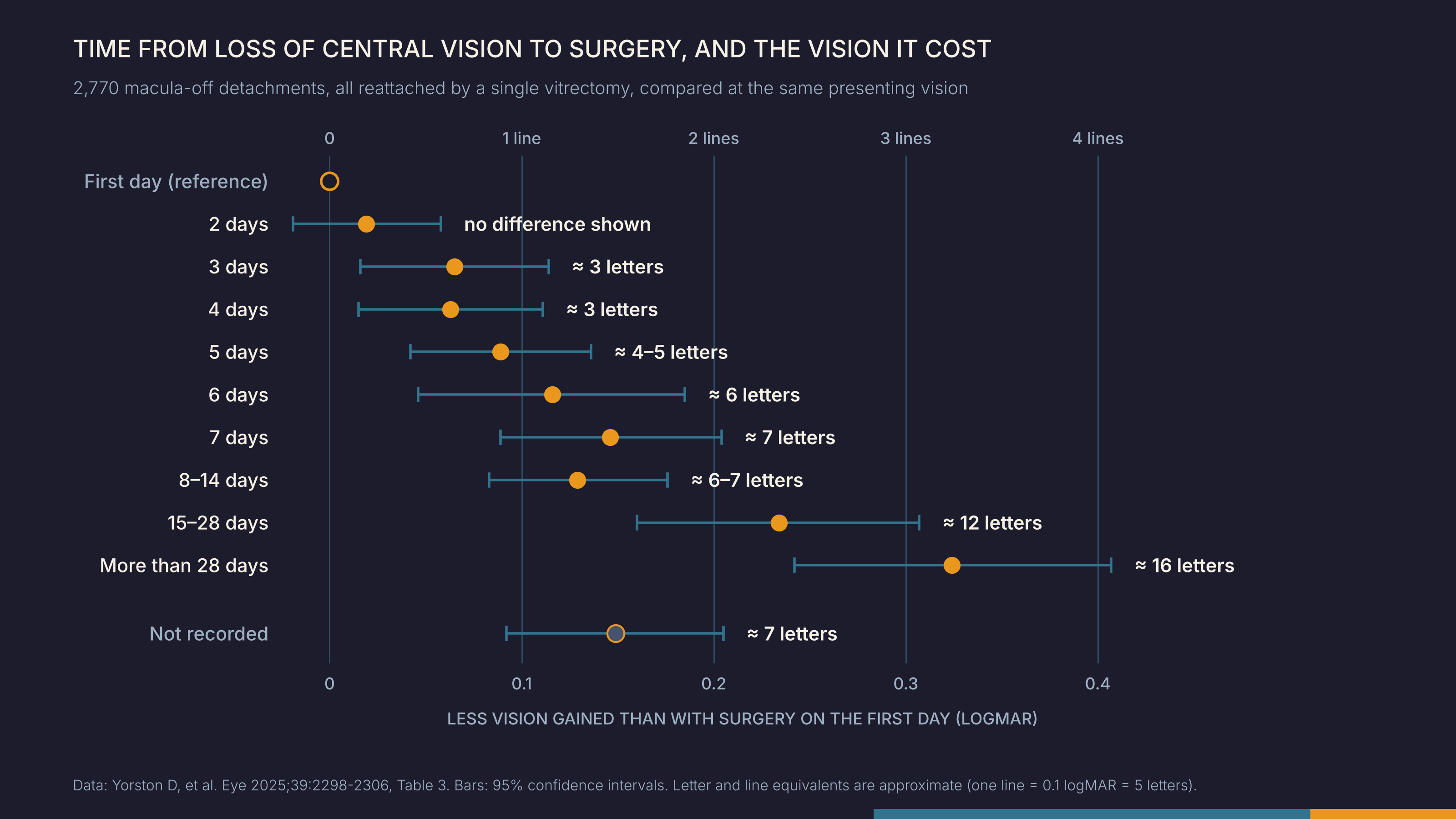
Among eyes that were all reattached and compared at the same presenting vision, the cost of delay rose from about three letters at three days to more than three lines beyond four weeks, with no point at which waiting stopped costing. A Japanese registry of 719 eyes is often read as showing such a point. Miyake and colleagues found better six-month acuity with surgery within two days, and within three, than with later surgery, and reported no significant difference at the cut-offs of four and five days. Their estimates at those two cut-offs, 0.063 and 0.061 logMAR, sit close to the 0.076 they reported as significant at three days; they fell short because the study set its threshold at P < 0.01, and their P values were 0.011 and 0.018. At each step the estimated effect stays near three letters. The registry also included every operated eye, whether or not the retina stayed attached, and it remains observational: the authors name residual confounding by indication as their first limitation.
All of this depends on a number we rarely question: the day central vision went. It comes from the patient's memory. In a Dutch series reported in a letter, 14 of 35 patients with a macula-off detachment could not give a time at all, and when patients whose macula was still attached were asked the same questions, between 15 and 40 per cent of their answers sounded as if it had come off. In the British database the duration was missing for one eye in four, and those eyes did about as badly as eyes that had waited a week.
The clock we schedule by is read from the patient's memory, and in a quarter of the largest database it was missing altogether. There is something we can measure instead. In a prospective series from Munich, 61 patients with a macula-off detachment of a week or less were treated by vitrectomy and gas, and all were pseudophakic by the end. Optical coherence tomography on the day of surgery measured the height of fluid under the fovea. At three months, eyes with less than 100 µm of subfoveal fluid reached a mean of 0.04 logMAR (about 0.9 in decimal acuity), and eyes with more than 1,500 µm reached 0.28 (about 0.5). Within that week, the number of days added nothing measurable once height was known. The series is small, with four and seven patients in its two lowest groups and three months of follow-up, and it excluded patients who could not date their loss of vision, who are exactly the patients the clock fails. It is too small to detect an effect of three letters. The authors' own conclusion is that surgery should not wait, because the height can keep rising.
The reattachment rate sees none of this. Every eye in the British analysis was a success by that measure. What separated them was time, which we know only roughly, and the height of the fovea, which can be measured on the morning of surgery and is not even a field in the largest database. If those are the two things still open to us, and each day of the first week appears to count, what is the macula-off on our next routine list waiting for?
References
- van Bussel EM, van der Valk R, Bijlsma WR, La Heij EC. Impact of duration of macula-off retinal detachment on visual outcome: a systematic review and meta-analysis of literature. Retina. 2014;34(10):1917-25. doi:10.1097/IAE.0000000000000296.
- Baumann C, Kaye SB, Steel DH. Reversing the paradigm on the urgency of acute retinal detachments defined by their foveal status: when off may be more urgent than on. BMJ Open Ophthalmol. 2024;9:e001668. doi:10.1136/bmjophth-2024-001668.
- Cook B, Lewis GP, Fisher SK, Adler R. Apoptotic photoreceptor degeneration in experimental retinal detachment. Invest Ophthalmol Vis Sci. 1995;36(6):990-6.
- Sothivannan A, Eshtiaghi A, Dhoot AS, Popovic MM, Garg SJ, Kertes PJ, Muni RH. Impact of the time to surgery on visual outcomes for rhegmatogenous retinal detachment repair: a meta-analysis. Am J Ophthalmol. 2022;244:19-29. doi:10.1016/j.ajo.2022.07.022.
- Yorston D, Donachie PHJ, Laidlaw DA, Steel DH, Williamson TH, et al. Predicting post-surgery change in visual acuity after successful repair of macula-off retinal detachments: findings from a large prospective UK cohort study. Eye (Lond). 2025;39:2298-306. doi:10.1038/s41433-025-03752-y.
- Miyake M, Nakao S, Morino K, Yasukura S, Mori Y, Ishihara K, et al. Effect of duration of macular detachment on visual prognosis after surgery for macula-off retinal detachment: Japan-Retinal Detachment Registry. Ophthalmol Retina. 2023;7(5):375-82. doi:10.1016/j.oret.2023.01.014.
- Ng H, La Heij EC, van Meurs JC. The duration of macular detachment in retinal detachment is difficult to determine. Acta Ophthalmol. 2020;98:e396-7. doi:10.1111/aos.14294.
- Baumann C, Almarzooqi A, Johannigmann-Malek N, Maier M, Kaye S. Importance of subfoveal fluid height on visual outcome in macula-off retinal detachments. Br J Ophthalmol. 2023;107:1467-71. doi:10.1136/bjophthalmol-2022-321604.