The Number That Was Not There
Two numbers govern how we follow an acute posterior vitreous detachment, and both vary across an order of magnitude. One of them varies partly because a figure in the most recent summary of this literature does not exist in the paper it is attributed to


A woman of sixty-four describes three days of flashes in the temporal field of her right eye and a new floater she compares to a tadpole. She has no relevant ocular history, no myopia worth the name, and she is phakic. Her acuity is unchanged, there is a Weiss ring, the anterior vitreous is clear of pigment, and there is no vitreous or retinal haemorrhage. Indirect ophthalmoscopy with scleral indentation through three hundred and sixty degrees shows no break and no lattice. She asks whether she should worry, and then a second question that matters more to us. When do you see her again?
The honest answer is that it depends on which paper you read last.
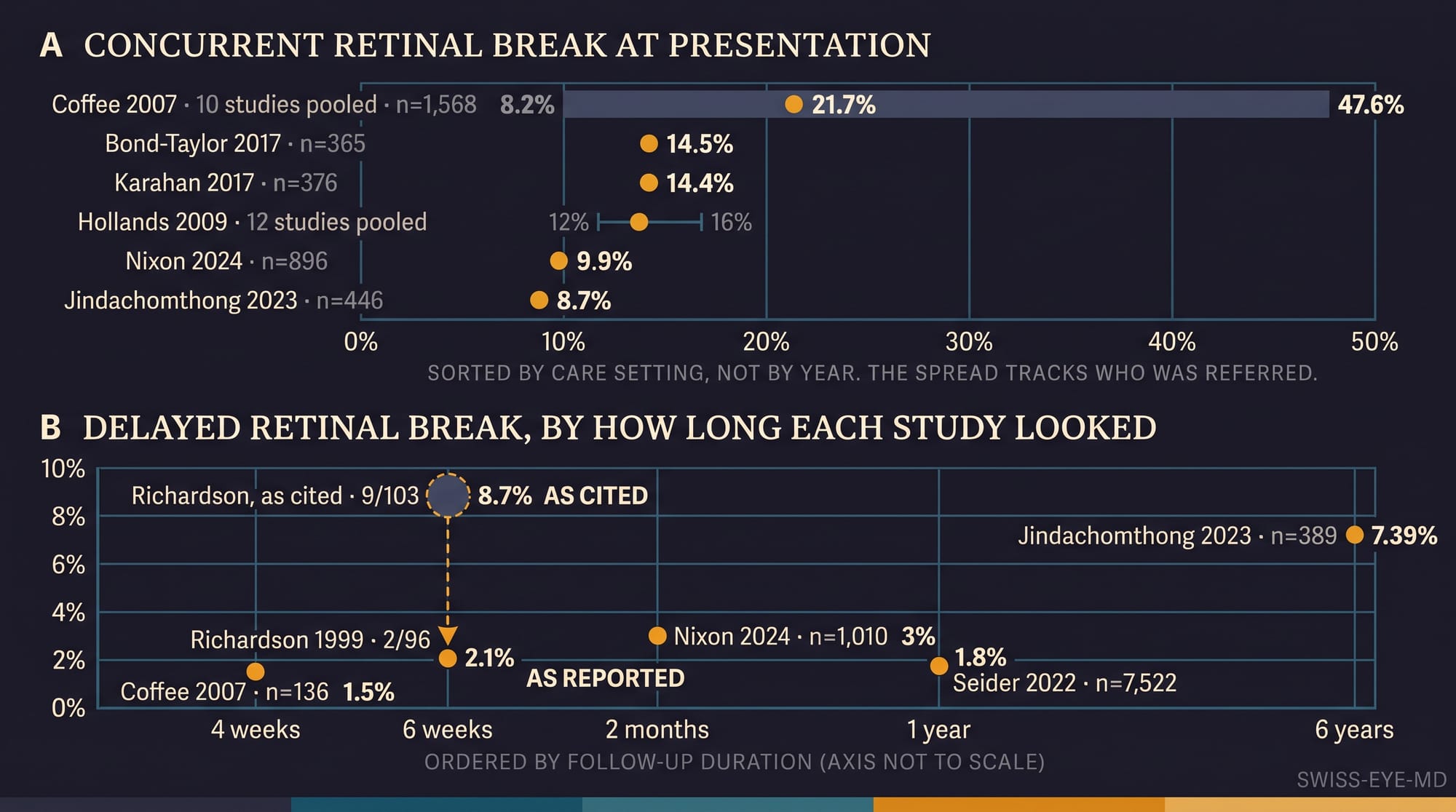
Across the published literature, somewhere between 5% and 48% of eyes presenting with an acute symptomatic posterior vitreous detachment are found to have at least one concurrent retinal break. That is not a confidence interval. It is the spread between individual studies, close to a tenfold difference in the thing we are counselling her about. The meta-analysis that pooled ten of these cohorts, 1,568 patients in all, reported 21.7%, with its component studies running from 8.2% to 47.6%.
The spread reflects who walked through each door rather than anything about the vitreous. A retina-only referral practice sees a population already filtered by an optometrist who suspected something; a community clinic seeing self-referred floaters sees a different disease prevalence under the same name. The authors of that meta-analysis attributed the variability to referral patterns and to the availability of vitreoretinal specialists at each study site, and there is no reason to look further.
The second number behaves the same way, and this is where it becomes interesting. Among eyes with no break at presentation, the most recent summary of this literature, a 2023 cohort study by Jindachomthong and colleagues, gives the delayed tear rate as 1.5% to 8.6%. The 1.5% comes from 136 patients re-examined within four weeks. The highest value in the table behind that range is 8.7%, attributed to a 1999 series from Birmingham, as nine delayed tears in 103 patients.
Open that paper and the nine is there. It reads: nine patients were referred for indirect laser photocoagulation following their first visit to the posterior vitreous detachment clinic. Six round holes, two horseshoe tears, one area of lattice with holes. Those nine are the tears found at presentation. The paper's own figure for the second visit, five weeks later, is two, in ninety-six patients given appointments, and it says of both that a vitreous haemorrhage at the first visit had obscured the retinal view. The denominator of 103 appears nowhere in it. Its own percentage for the nine is 8.4%, and that is nine over the 107 patients it actually saw
The highest delayed-tear figure in that table is a first-visit figure that has been read as a follow-up figure, and the paper it comes from concludes the opposite of what that reading implies. Its abstract closes on exactly that point: if no retinal breaks are detected by thorough examination at presentation, further reviews, in the absence of increasing symptoms, may not be necessary. And the two patients in whom a break was found at the second visit appear to be the two the summary table removes from its denominator, on the grounds that a vitreous haemorrhage had obscured a tear already present. The discrepancy has been communicated to the corresponding author of the 2023 study.
None of this makes the delayed risk zero, and the largest dataset we have says so plainly. In the American Academy's IRIS Registry, 434,046 eyes met the criteria for an acute posterior vitreous detachment, and 10,518 of them, 2.42%, were found to have a delayed or missed retinal break or detachment within the following year. In the eyes documented with vitreous haemorrhage and nothing else, 657 of them, the median time to a break was fourteen days. A registry of that size cannot tell us what any individual eye will do, and it carries the coding limitations its authors set out at length, but it does establish that the tail is real and that it is not evenly distributed.
The point is narrower and more uncomfortable than the numbers themselves. Two of the figures we quote to patients are a pooled estimate whose spread is driven by catchment, and a range whose highest tabulated value, in the most recent summary of this literature, does not survive a reading of the paper it is drawn from.
So what does the guidance say. The American Academy's Preferred Practice Pattern sets four to six weeks for a symptomatic detachment with no break and no high-risk feature, one to two weeks where there is retinal haemorrhage, and weekly review until resolution where there is vitreous haemorrhage. Europe has no equivalent document of comparable reach, and most of us are working from the American pattern, from local custom, or from what our training left us with.
That is a real stratification, and it runs on a single axis: blood, or no blood. It is silent on lattice degeneration, on a history in the fellow eye, and on how quickly she presented. It is also, by construction, a decision that can only be taken after someone has dilated her and indented her periphery, which means it does no work at the point where most of these patients first make contact, which is a telephone call to a clinic deciding who is seen tomorrow and who is seen in a fortnight.
Return to her. She has none of the features that would move her out of the default tier, and none of the reassurance a negative examination is often taken to provide, because a negative examination is a statement about today. The Birmingham series would discharge her. The six-year cohort found more than half of its delayed tears after the point at which she would have been discharged. Both are looking at the same eye.
The next entry asks what actually predicts a break, which is a harder question than the textbook account suggests, because the symptom we ask about most confidently is the one on which the evidence disagrees most sharply.
For now, the question she asked, reframed. If the interval we give her rests on figures we have not opened, what exactly is it resting on?
References
- Coffee RE, Westfall AC, Davis GH, Mieler WF, Holz ER. Symptomatic posterior vitreous detachment and the incidence of delayed retinal breaks: case series and meta-analysis. Am J Ophthalmol. 2007;144(3):409-13. doi:10.1016/j.ajo.2007.05.002.
- Richardson PS, Benson MT, Kirkby GR. The posterior vitreous detachment clinic: do new retinal breaks develop in the six weeks following an isolated symptomatic posterior vitreous detachment? Eye (Lond). 1999;13(Pt 2):237-40. doi:10.1038/eye.1999.58.
- Jindachomthong KK, Cabral H, Subramanian ML, et al. Incidence and risk factors for delayed retinal tears after an acute, symptomatic posterior vitreous detachment. Ophthalmol Retina. 2023;7(4):318-24. doi:10.1016/j.oret.2022.10.012.
- Kim SJ, Bailey ST, Kovach JL, et al. Posterior vitreous detachment, retinal breaks, and lattice degeneration preferred practice pattern. Ophthalmology. 2025;132(4):P163-P196. doi:10.1016/j.ophtha.2024.12.023.
- Hollands H, Johnson D, Brox AC, Almeida D, Simel DL, Sharma S. Acute-onset floaters and flashes: is this patient at risk for retinal detachment? JAMA. 2009;302(20):2243-9. doi:10.1001/jama.2009.1714.
- Vangipuram G, Li C, Li S, et al. Timing of delayed retinal pathology in patients presenting with acute posterior vitreous detachment in the IRIS Registry (Intelligent Research in Sight). Ophthalmol Retina. 2023;7(8):713-20. doi:10.1016/j.oret.2023.04.004.