The Atrophy Arc, End to End
The four-part Atrophy Arc in one place. Every advance, from defining the lesion to slowing it, is a growing command over an anatomical endpoint, while the patient's vision stays out of frame.

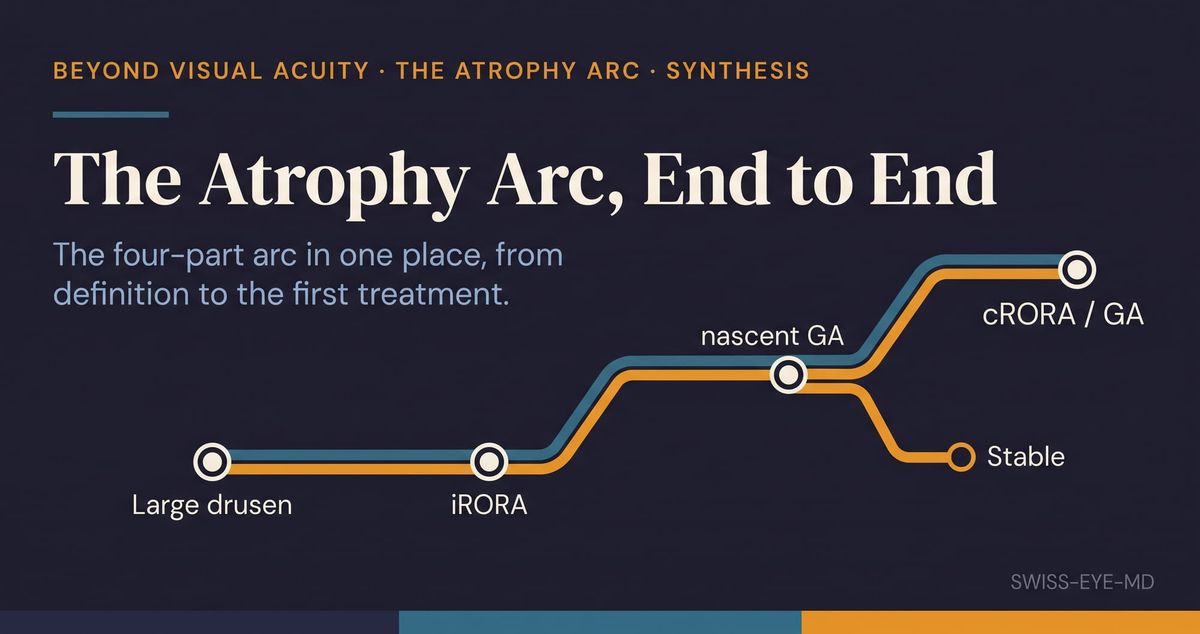
Four Wednesdays, four entries, one endpoint. This series has followed geographic atrophy from the moment it is defined on a scan to the moment a drug is offered against it, and along the way it kept circling the same quiet tension. Everything we learned to do with this disease, we learned to do to an image of it. Now that the arc is complete, that thread is worth pulling on.
The arc began by fixing what we were talking about. What we mean by atrophy settled on cRORA, complete RPE and outer retinal atrophy, as the endpoint: a column of hypertransmission into the choroid, a wide break in the RPE, and degeneration of the photoreceptors above it, each meeting a size threshold on OCT. That did a real service, because a field cannot study what it cannot name the same way twice. It is worth being clear about what kind of thing it named. cRORA is an anatomical description of an image. It records where tissue has been lost and how much, and it says nothing directly about what the eye can still see. Everything the arc did afterwards was built on that footing.
A definition is only worth what two readers can independently reproduce, and the second entry asked whether they can. Twelve readers grading the same scans agreed well on most of the OCT signs of early atrophy, which is reassuring for an endpoint meant to travel between reading centres. The awkward exception was the RPE edge, the attenuation and disruption that give cRORA its "complete", where agreement was weakest. The signs the endpoint is named after are the ones two experts are least sure they are seeing the same way. The composite call still held together, because the classification leans on the clearer signs beneath it. But the first crack in the surrogate showed here: some of what we count as the lesion growing is the reader moving.
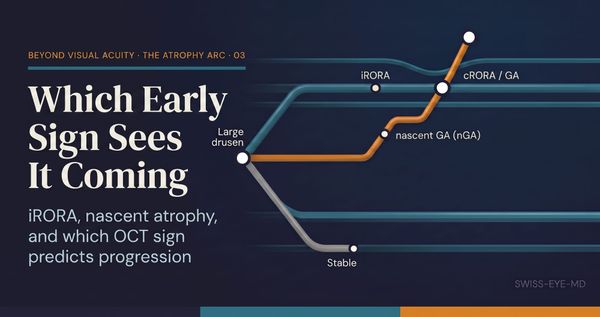
If we can define the lesion and mostly agree on it, the next thing worth having is warning. The third entry asked which early sign sees atrophy coming. iRORA, the consensus precursor, did raise the risk of progression. Followed over time, though, a different sign predicted it better: nascent geographic atrophy, the subsidence of the deeper retinal layers and the hyporeflective wedge within Henle's fibre layer, which largely absorbed iRORA's signal once both were tested together. The sign that best foretold the endpoint was the one two readers agreed on most readily, and it sits outside the definition of the precursor we had chosen to name. Prediction, like definition and agreement before it, was read off structure. The eye's future was inferred from the shape of its layers, never from a measure of sight.
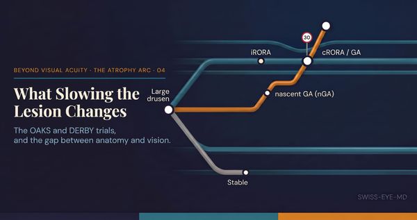
And then, for the first time, something to do about it. The fourth entry took up the first approved treatment, a complement inhibitor that slows the growth of the atrophic lesion. The effect is real and it is modest, and its shape is instructive. One phase 3 trial met its twelve-month primary endpoint. Its identically designed twin did not. Across both, the drug spared a rim of retinal tissue from disappearing, and across both, that spared tissue did not translate into vision the eye could use: letters read, reading speed, and the patient's own sense of function all moved no better than under sham. The treatment slows the map of the disease. Whether it changes the disease as the patient lives it is still unproven, and the harms are not nothing.
Set the four entries side by side and one thread runs the length of them. Every step of this arc, from defining the lesion to slowing it, is a growing command over an anatomical endpoint, and at no point did the thing that matters most to the patient, their vision, come into frame. The least reliable signs of the second entry, the structural predictor of the third, and the anatomic-without-functional benefit of the fourth are the same fact seen from different angles. What the arc settled is not small. We can now define the lesion, read it within known limits, name the early sign that best foretells it, and slow it once it has formed. Those are four genuine advances in how we handle geographic atrophy on a scan.
What it did not settle is the thing a patient would ask first. We have learned to move the lesion. We have not yet shown that moving the lesion moves the patient. The distance between the anatomy we can now influence and the sight we cannot yet promise is where this arc ends, honestly, and without closing it. If you are new here, the four entries read best in order, beginning with what we mean by atrophy. And if you have followed all four, the question they leave is the one worth carrying: when we slow a lesion we can measure, how confident should we be that we are helping the person whose sight it is taking?
References
- Sadda SR, et al. CAM Report 3. Ophthalmology. 2018;125(4):537-548.
- Wu Z, et al. CAM Report 6. Ophthalmology Retina. 2022;6(1):4-14.
- Wu Z, Goh KL, Hodgson LAB, Guymer RH. Ophthalmology. 2023;130(2):205-212.
- Heier JS, et al. OAKS and DERBY. Lancet. 2023;402:1434-1448.