What a Flash Is Worth
Six research groups have asked whether photopsia predicts a retinal tear. They disagree, and one of them finds it mildly protective. The findings that hold are the ones we ask about least confidently.

The last entry ended on the interval and what it rests on. Underneath that question sits a simpler one that ought to have been settled decades ago. When a patient reports flashing lights, what have we learned?
Start with the number the whole calculation hangs on. Among patients with an acute posterior vitreous detachment who are self-referred or referred to an ophthalmologist, the summary prevalence of a retinal tear is 14%, with a confidence interval from 12% to 16%. That is the pre-test probability, and the meta-analysis that produced it notes that every study in it came from an ophthalmology clinic, and that in primary care the figure may well be lower.
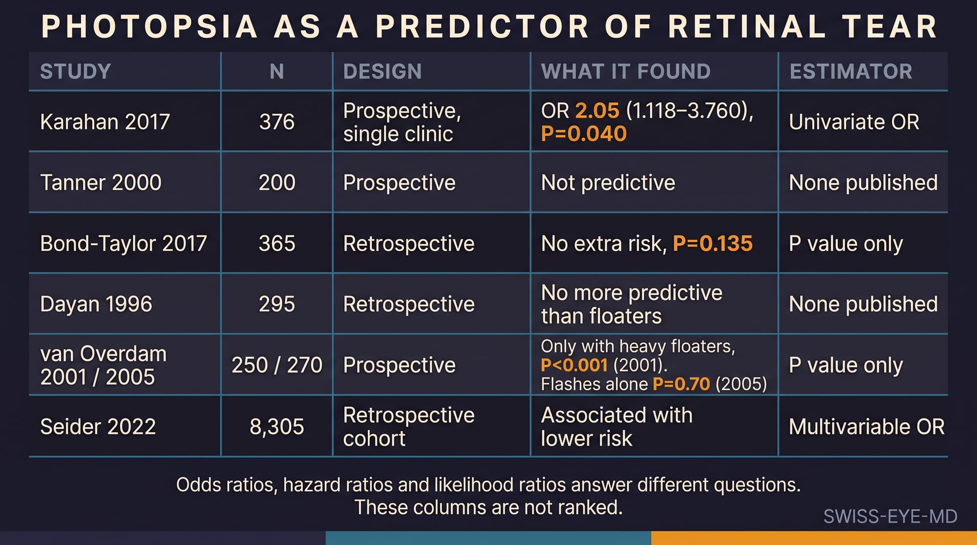
Now the flash. A prospective series of 376 consecutive patients found photopsia associated with a retinal tear at an odds ratio of 2.05, confidence interval 1.118 to 3.760, on univariate analysis. A series of 200 patients concluded that it was not possible on the basis of symptoms alone to determine which patients had a break, and found that flashes and floaters together carried no significantly higher chance than floaters alone. A Swedish cohort of 365 reported that symptoms of flashes did not constitute an extra risk, at P=0.135. A Dutch group found flashes predictive in their 2001 series only in combination with a heavy floater burden, more than ten floaters or a cloud or curtain, and in their 2005 series flashes reached a P value of 0.70, the authors noting that the small sample size of the earlier study may explain the difference. A retrospective review of 295 patients found isolated photopsia no more predictive than floaters. And a retrospective cohort of 8,305 patients at Kaiser Permanente found photopsias associated with a lower risk of a complicated posterior vitreous detachment, at an odds ratio of 0.76, offering as a possible explanation that patients with significant vision loss do not report what may feel like a milder symptom.
Six research groups have asked the same question of the same symptom and produced an odds ratio above two, three null results, one failed internal replication, and one inverse association. A symptom that behaves like that across seven datasets is not a weak predictor. It is an unstable one, and the instability is worth more diagnostic attention than the point estimates.
Against that, look at what holds. Subjective reduction in vision carries a positive likelihood ratio of 5.0, confidence interval 3.1 to 8.1, from a single series of 295 patients in which sixty-seven per cent of those reporting visual loss had a break or a detachment, against eighteen to twenty-three per cent in every other symptom group. Vitreous haemorrhage at examination carries a summary positive likelihood ratio of 10, confidence interval 5.1 to 20, from eleven studies. Vitreous pigment carries a summary of 44, with a confidence interval from 2.3 to 852 that tells you how little that point estimate should be trusted.
The arithmetic follows. Starting from 14%, a subjective reduction in vision raises the probability of a tear to 45%, with a confidence interval of 34% to 57%. Vitreous haemorrhage raises it to 62%, from 45% to 77%. Those are the meta-analysis's own published post-test figures, not a calculation done here, and both are decision-changing in a way that no reported effect for photopsia is.
There is a third finding of that kind, and it is the closest anything in this literature comes to a stratification rule. In 8,305 patients seen across a large integrated health system, of whom 7,522 had an initially uncomplicated detachment, three features present at the first visit separated the groups cleanly. Vitreous haemorrhage, lattice degeneration, or a history of a tear or a detachment in the fellow eye: patients with at least one of the three had a 12.4% rate of a late tear or detachment within a year, and patients with none of them had 0.7%. None of those three is a symptom. Two of them require a dilated examination, and the third requires knowing what happened to the other eye.
Two cautions belong with this, and they are the reason the picture stays messier than the numbers suggest.
The first is that these estimates are not commensurable. Odds ratios, hazard ratios and likelihood ratios answer different questions, and lining them up by magnitude to produce a hierarchy of risk factors is an error that looks like rigour. An odds ratio of 12.01 for constant blurred vision or a curtain in a multivariable telephone model, a hazard ratio of 6.48 for lattice degeneration against a time-to-event endpoint, and a likelihood ratio of 10 for vitreous haemorrhage against a same-visit endpoint cannot be ranked against one another, however tempting the column of figures is.
The second caution is that the sign most of us trust to close the question confirms better than it excludes. The Shafer sign is usually highly specific: 100% in three of the four series in that meta-analysis and 98.2% in a later one, although the largest of the four, 589 patients, reported 68%. Its sensitivity is another matter, reported at 23.8% in one series built on five true-positive eyes, 63% in another, 79%, 92% and 94% in others, with a summary negative likelihood ratio of 0.23 and a confidence interval reaching 0.43. A clear anterior vitreous in an eye with a fresh detachment means considerably less than its reputation suggests. And in a prospective study of 1,010 community referrals, a Weiss ring was absent in thirty-two per cent of the eyes that had a retinal tear, while fifteen per cent of tears were visible only on indented indirect ophthalmoscopy and not on slit-lamp biomicroscopy.
Which leaves the encounter in an awkward shape. The symptom the patient volunteers first and describes most vividly is the one the evidence cannot agree on. The findings that move the probability are the ones that require a dilated pupil, a slit lamp, and an examiner willing to indent. And the sign we use to reassure ourselves can miss a substantial share of what we are looking for.
If photopsia has earned an odds ratio of 2.05 in one cohort and a protective association in another, what are we actually doing when we ask about it?
References
- Hollands H, Johnson D, Brox AC, Almeida D, Simel DL, Sharma S. Acute-onset floaters and flashes: is this patient at risk for retinal detachment? JAMA. 2009;302(20):2243-9. doi:10.1001/jama.2009.1714.
- Karahan E, Karti O, Er D, et al. Risk factors for multiple retinal tears in patients with acute posterior vitreous detachment. Int Ophthalmol. 2018;38(1):257-63. doi:10.1007/s10792-017-0455-0.
- Tanner V, Harle D, Tan J, Foote B, Williamson TH, Chignell AH. Acute posterior vitreous detachment: the predictive value of vitreous pigment and symptomatology. Br J Ophthalmol. 2000;84(11):1264-8. doi:10.1136/bjo.84.11.1264.
- Bond-Taylor M, Jakobsson G, Zetterberg M. Posterior vitreous detachment: prevalence of and risk factors for retinal tears. Clin Ophthalmol. 2017;11:1689-95. doi:10.2147/OPTH.S143898.
- van Overdam KA, Bettink-Remeijer MW, Mulder PG, van Meurs JC. Symptoms predictive for the later development of retinal breaks. Arch Ophthalmol. 2001;119(10):1483-6. doi:10.1001/archopht.119.10.1483.
- van Overdam KA, Bettink-Remeijer MW, Klaver CC, Mulder PG, Moll AC, van Meurs JC. Symptoms and findings predictive for the development of new retinal breaks. Arch Ophthalmol. 2005;123(4):479-84. doi:10.1001/archopht.123.4.479.
- Dayan MR, Jayamanne DGR, Andrews RM, Griffiths PG. Flashes and floaters as predictors of vitreoretinal pathology: is follow-up necessary for posterior vitreous detachment? Eye (Lond). 1996;10(Pt 4):456-8. doi:10.1038/eye.1996.100.
- Seider MI, Conell C, Melles RB. Complications of acute posterior vitreous detachment. Ophthalmology. 2022;129(1):67-72. doi:10.1016/j.ophtha.2021.07.020.
- Balikov DA, Zhou Y, Miller JML. A telephone triage system for patients calling with symptoms of a posterior vitreous detachment. Ophthalmol Retina. 2023;7(6):516-26. doi:10.1016/j.oret.2023.01.003.
- Jindachomthong KK, Cabral H, Subramanian ML, et al. Incidence and risk factors for delayed retinal tears after an acute, symptomatic posterior vitreous detachment. Ophthalmol Retina. 2023;7(4):318-24. doi:10.1016/j.oret.2022.10.012.
- Oh JH, Oh J, Roh HC. Vitreous hyper-reflective dots in optical coherence tomography and retinal tear in patients with acute posterior vitreous detachment. Curr Eye Res. 2017;42(8):1179-84. doi:10.1080/02713683.2017.1289226.
- Nixon TRW, Davie RL, Snead MP. Posterior vitreous detachment and retinal tear: a prospective study of community referrals. Eye (Lond). 2024;38:786-91. doi:10.1038/s41433-023-02779-3.