What Slowing the Lesion Changes
The first approved treatment for geographic atrophy slows the lesion on OCT in OAKS and DERBY. Read closely: real, partial, and not yet shown to protect sight.

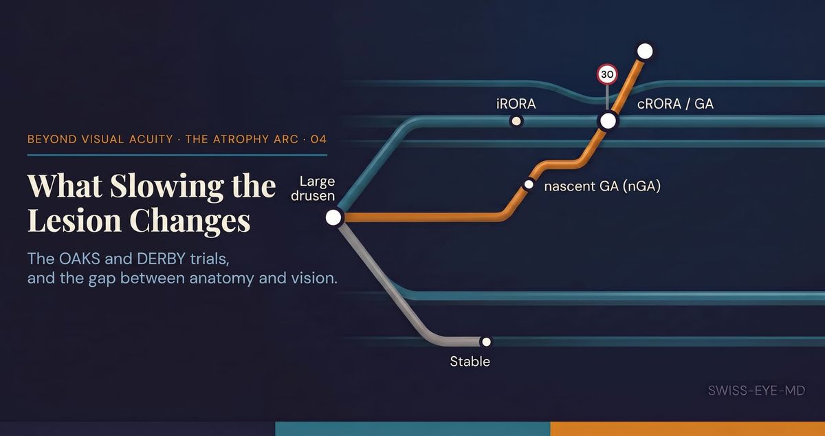
The previous entry ended on a deferral. Having asked which early sign best anticipates progression, it set aside the larger question for this one: whether acting on the lesion once it is established changes the course of the disease. For most of the history of geographic atrophy that question had no answer to test, because there was nothing approved to give. That has changed. Two phase 3 programmes, OAKS and DERBY, put the first complement inhibitor against the endpoint this arc has spent three entries defining, agreeing on, and learning to predict. What they show is worth reading closely, and reading honestly.
OAKS and DERBY were two identically designed trials of intravitreal pegcetacoplan, a C3 complement inhibitor, given either monthly or every other month against a sham injection, in eyes with geographic atrophy secondary to age-related macular degeneration. The primary endpoint in both was the change from baseline to month 12 in the total area of geographic atrophy lesions, measured on fundus autofluorescence. It is worth pausing on what that endpoint is. It is an anatomical quantity, the square millimetres of atrophy on an autofluorescence image and the rate at which they enlarge. It measures the lesion. It does not measure the person reading a chart. The whole of what follows turns on holding those two things apart.
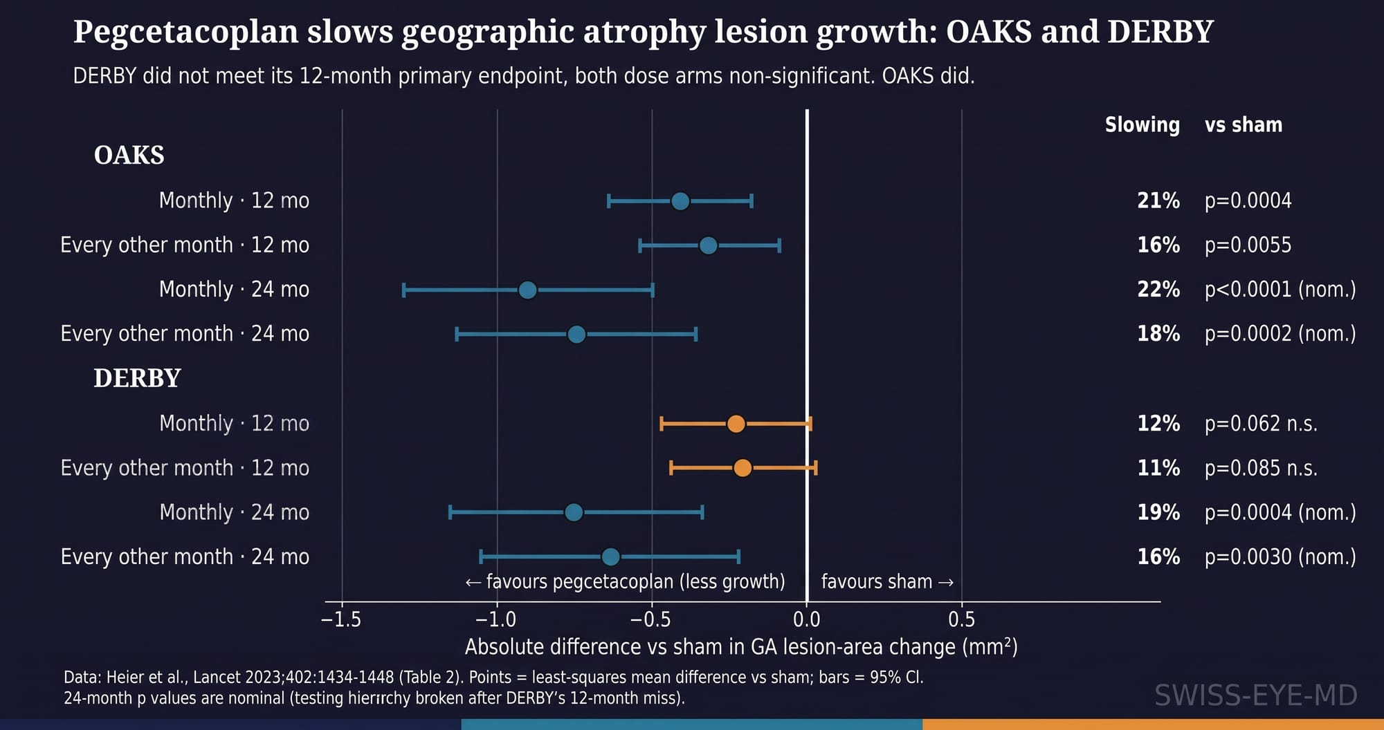
The two trials did not agree. In OAKS, pegcetacoplan slowed lesion growth at 12 months by 21% given monthly and 16% every other month, both significant against sham. In DERBY, the same drug on the same schedule slowed growth by 12% and 11%, and neither reached significance (p=0·062 and p=0·085). One pivotal trial met its primary endpoint and its twin did not. The first pharmacological effect ever shown on geographic atrophy is therefore real, and it is also partial and slow. By 24 months both trials showed a wider separation from sham, but those later p values are nominal: once DERBY missed at month 12 the prespecified testing hierarchy was broken, so nothing measured afterwards carries confirmatory weight. The honest reading is a drug with a genuine biological effect that one of its two identical trials could not confirm on the endpoint that mattered most.
The effect also accumulates rather than arrives. A piecewise analysis of the combined studies put the growth difference near 16% across the first year and larger, close to 30% in the monthly arm, only in the final six months. Slowing of this kind does not reverse atrophy and does not restore a retinal pigment epithelium or the photoreceptors that have already gone. It reduces the rate at which the lesion expands into tissue that is still intact. Over two years the average tissue spared is between about 0·6 and 0·9 mm². Whether that is worth an injection every month or two is a question the anatomy alone cannot answer.
That question falls to visual function, and here the trials are quiet. Across the combined studies there was no significant difference from sham in best-corrected visual acuity at 24 months, and the treated eyes were, if anything, a fraction of a letter worse. The prespecified secondary endpoints of maximum reading speed, functional reading independence and microperimetric sensitivity showed no significant benefit either. One narrow signal survived, a reduction in new scotomatous points within the junctional zone bordering the lesion, and even that was significant only in the every-other-month arm. We can slow the lesion, and these trials have not shown that slowing it changes what the patient sees.The investigators are candid about this in their own discussion, observing that the available functional measures are subjective and variable while the anatomical endpoint is objective and high in resolution. That is a fair defence of the endpoint. It is not a demonstration of benefit to the patient.
Against an unproven functional benefit sit real risks, and they are of the kind that threaten the vision the treatment is meant to protect. New-onset neovascular AMD in the treated eye reached 11% to 13% of monthly-treated eyes and 6% to 8% of those treated every other month by 24 months, against 2% to 4% on sham. Turning a slowly atrophic eye into a wet one carries its own burden of injections and its own risk to central vision. Intraocular inflammation occurred in up to 5% of monthly-treated eyes, most of it mild, and three eyes, all in the monthly arm, developed ischaemic optic neuropathy. The trials recorded no retinal vasculitis of any kind. The occlusive retinal vasculitis that later attached itself to this drug, and the regulatory divergence that followed in Europe, sit outside these data; I have set out that safety and regulatory picture separately in an earlier piece on complement inhibition. Within the trials themselves, the harm that recurs is conversion to neovascular disease, and it tracks with dose.
So the established-lesion decision is genuinely difficult, and the trial itself points to where the balance tips. In the prespecified subgroup analysis the effect was largest in non-subfoveal lesions, where growth at 24 months was slowed by around a quarter, and smallest where the atrophy was already subfoveal. The eyes with the most to gain are those whose lesion is advancing toward a fovea it has not yet reached, where buying time has a clear meaning. The eyes with the least to gain are those where the central damage is already done. Most patients present somewhere in between, in the territory where a slow anatomical benefit of unproven functional value is weighed against a real risk of neovascular conversion, in a disease whose untreated course is itself slow.
This closes the arc where it began. We set out to fix what we mean by atrophy, to agree on how to read it, and to learn which early sign foretells it; the treatment era now lets us slow the very lesion those three entries defined. Slowing an anatomical endpoint is the thing that has been proven, and protecting sight is the thing that has not, and the distance between them is the whole of what this arc leaves unsettled. A synthesis of the four entries follows, drawing together what has been established and what stays open. Before it, one question is worth sitting with. If the measure we can move is not yet the measure the patient feels, how much certainty do we owe them about that difference before we reach for the syringe?
References
- Heier JS, Lad EM, Holz FG, et al. Pegcetacoplan for geographic atrophy secondary to age-related macular degeneration (OAKS and DERBY): two multicentre, randomised, double-masked, sham-controlled, phase 3 trials. Lancet 2023;402:1434-1448.