What We Mean by Atrophy
The Atrophy Arc opens by defining its endpoint: cRORA, complete RPE and outer retinal atrophy, and the four consensus criteria that mark it on OCT


This series paused in July on one line: that the hyperreflective foci matter less for what they are than for the atrophy they anticipate, defined layer by layer. That line assumed something it never spelled out: what atrophy on OCT actually is. Before we can ask how imaging anticipates atrophy, we have to pin down what is being anticipated.
The framework comes from the Classification of Atrophy Meetings, an international consensus set up to standardise how atrophy in age-related macular degeneration is named and identified on OCT. Two of its decisions matter here. The first is that OCT was made the base imaging method for diagnosing and staging atrophy, with fundus autofluorescence, near-infrared reflectance and colour imaging kept in a complementary, confirmatory role. The second is that atrophy was treated as a graded process, moving through stages toward an end state, rather than a condition that is simply present or absent. Four terms were proposed for that spectrum; the one this entry defines is the complete form, complete RPE and outer retinal atrophy, or cRORA. In eyes without neovascularisation it corresponds to what we have long called geographic atrophy, a term the consensus kept and restricted to exactly that setting. Colour fundus photography identified that atrophy for decades; OCT was chosen because it shows the outer-retinal and pigment-epithelial change beneath it that photography cannot. The other three terms fill in the rest: an incomplete counterpart to cRORA, and two forms in which the outer retina atrophies while the pigment epithelium is still spared. cRORA is the complete, RPE-involving end of that spectrum; the incomplete stages before it are what the rest of this arc is about.
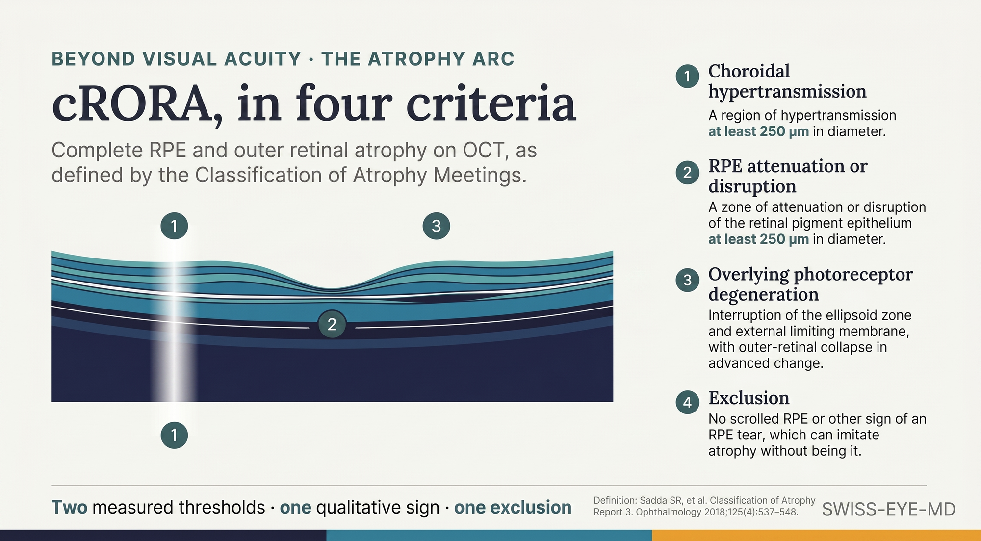
cRORA is defined by explicit criteria, not by overall impression. The consensus proposed four. First, a region of hypertransmission of at least 250 μm in diameter, the bright column that appears beneath the retinal pigment epithelium where light passes through tissue that no longer absorbs it. Second, a zone of attenuation or disruption of the RPE of at least 250 μm in diameter. Third, evidence of overlying photoreceptor degeneration, seen as interruption of the ellipsoid zone and the external limiting membrane and, in more advanced change, subsidence of the inner nuclear and outer plexiform layers as the outer retina collapses inward. Fourth, an exclusion: the absence of scrolled RPE or other signs of an RPE tear, which can imitate atrophy without being it. The two size thresholds keep small or incidental change from being called atrophy, and the exclusion guards against a tear that can look the same. The definition, then, is two measured thresholds, one qualitative sign, and one thing that must be absent.
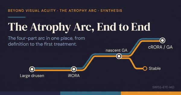
The same consensus named an incomplete form, iRORA, and it is worth naming here even though this entry does not set out its full criteria. In iRORA the hypertransmission is present but discontinuous, the RPE band is irregular or interrupted rather than lost, and the criteria for cRORA are not yet met. It sits earlier on the same trajectory, an atrophy that has begun without completing. Its edges are harder to call than those of the complete form, which is part of why it is named on its own. That earlier phenotype is where this arc turns, because the question of anticipation lives between the incomplete form and the complete one. A later entry takes up its formal definition and the evidence on how often, and how fast, it crosses over. For now it stays a described precursor, one whose predictive weight this arc will test rather than assume.
Two consequences follow from defining the endpoint this precisely, and both point beyond this entry. The first is a problem of agreement. A threshold of 250 μm, an interrupted ellipsoid zone, a judgement that the RPE is disrupted rather than merely thinned: each of these has to be read off a real scan by a real observer, and a definition is worth only as much as the consistency with which two readers apply it to the same image. That consistency matters beyond the reading centre: it is what lets a diagnosis reached in one clinic mean the same thing in another, and an endpoint measured at one trial site stay comparable with the next. Whether experienced graders in fact agree that these criteria are met, and where they part company, is the subject of the next entry. The second consequence is that the endpoint has outgrown its descriptive role: it is now how trials measure the disease, and the reference against which the first approved treatments state their effect, taken up in the closing entries of the arc on their own terms.
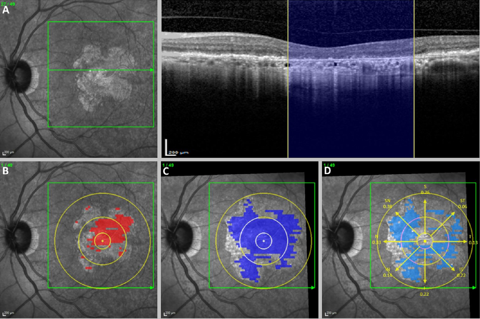
One rule from the previous arc carries over here. The criteria for cRORA describe an imaging phenotype, a pattern of hypertransmission, pigment epithelial loss and photoreceptor change. They do not describe a directly observed cellular event. The correspondence between that phenotype and the underlying histology is well supported, and the consensus was built partly on paired imaging and histologic review, yet it remains an inference drawn from light returning through tissue. The consensus was candid about this, pairing each term with a histologic candidate, a best current reading of the tissue beneath the OCT appearance, and noting that longitudinal data would be needed to validate the risk of vision loss these terms imply. The prognostic weight these features carry, and increasingly the therapeutic weight, rests on that imaging signature. It is a defensible place to rest it, as long as the inference is called an inference and not quietly upgraded to a proven mechanism.
Defining the endpoint is the least exciting part of an arc about anticipation, and the part the rest of it leans on. How reliably the endpoint is recognised, how its incomplete form predicts the complete one, and whether treatment alters either: all of it depends on the definition holding still while those questions are asked. So the question to put to colleagues, before any predictive one, is simple. Shown the same B-scan and the same four criteria, how often would two of us agree that atrophy, in the complete sense defined here, is actually there?
References:
- Sadda SR, et al. Consensus Definition for Atrophy Associated with Age-Related Macular Degeneration on OCT: Classification of Atrophy Report 3. Ophthalmology. 2018;125(4):537-548.
- Shmueli O, Yehuda R, Szeskin A, Joskowicz L, Levy J. Progression of cRORA (Complete RPE and Outer Retinal Atrophy) in Dry Age-Related Macular Degeneration Measured Using SD-OCT. Transl Vis Sci Technol. 2022;11(1):19.